Author: Denis Avetisyan
A new deep learning model uses the power of temporal imaging to forecast long-term Alzheimer’s disease progression, even with irregularly spaced scan data.

Researchers demonstrate improved prediction accuracy by modeling uncertainty with a Normal Inverse Gamma distribution and extracting key temporal features from brain imaging.
Predicting the long-term progression of Alzheimer’s Disease remains challenging, particularly when faced with irregularly spaced patient imaging data. This limitation motivates the research presented in ‘Long-Term Alzheimers Disease Prediction: A Novel Image Generation Method Using Temporal Parameter Estimation with Normal Inverse Gamma Distribution on Uneven Time Series’, which introduces a novel deep learning model, T-NIG, to forecast disease progression by explicitly modeling temporal dependencies within brain imaging sequences. By leveraging the Normal Inverse Gamma distribution and uncertainty estimation, T-NIG demonstrates state-of-the-art performance in maintaining disease-related characteristics even with inconsistent data. Could this approach pave the way for earlier and more accurate diagnoses, ultimately improving patient outcomes?
Whispers of Future Brains: Forecasting Neurological Change
The ability to forecast longitudinal brain changes holds immense promise for transforming the landscape of neurodegenerative disease management. Precise prediction isn’t merely about identifying illness; it’s about pinpointing vulnerability before symptoms manifest, potentially allowing for preventative interventions and dramatically slowing disease progression. For conditions like Alzheimer’s and Parkinson’s, where neuronal damage often begins decades before clinical presentation, this predictive capability is especially crucial. Furthermore, tracking these changes over time provides a more sensitive measure of therapeutic efficacy than relying solely on static assessments, enabling clinicians to personalize treatment strategies and optimize patient care. Ultimately, improved forecasting will shift the focus from reactive treatment to proactive, preventative healthcare, significantly improving outcomes and quality of life for those at risk.
Conventional analyses of brain imaging data frequently treat each scan as a static snapshot, failing to fully account for the continuous, evolving nature of neurological processes. This approach overlooks crucial information embedded within the temporal progression of brain changes-the subtle shifts occurring over months or years. Consequently, forecasts of disease progression, or individual responses to treatment, often prove inaccurate. The brain isn’t simply different at two points in time; how it changes-its velocity and trajectory-is equally important. Standard statistical models struggle to represent this dynamic interplay, leading to imprecise predictions and hindering the development of truly personalized medicine for neurodegenerative conditions. Capturing these complex temporal dynamics requires innovative methodologies capable of modeling brain states not as isolated events, but as points along a continuous, potentially non-linear, path.
Predicting the trajectory of brain changes demands more than simply identifying trends; it necessitates a nuanced understanding of both the data’s limitations and the model’s predictive capabilities. Brain imaging data, by its very nature, contains inherent noise – random fluctuations arising from biological variability and measurement error. A robust temporal model doesn’t just smooth over this noise, but explicitly accounts for it, acknowledging that any prediction is probabilistic, not deterministic. Crucially, the model itself possesses uncertainty; its forecasts are not absolute truths, but rather estimations based on past observations. Therefore, effective modeling incorporates a quantification of this epistemic uncertainty – a measure of how confident the model is in its own predictions – providing a more realistic and reliable assessment of future brain states. This approach moves beyond point predictions, offering a distribution of possible outcomes, allowing clinicians to better understand the range of potential disease progression and tailor interventions accordingly.
The T-NIG Model: A Probabilistic Dance with Time
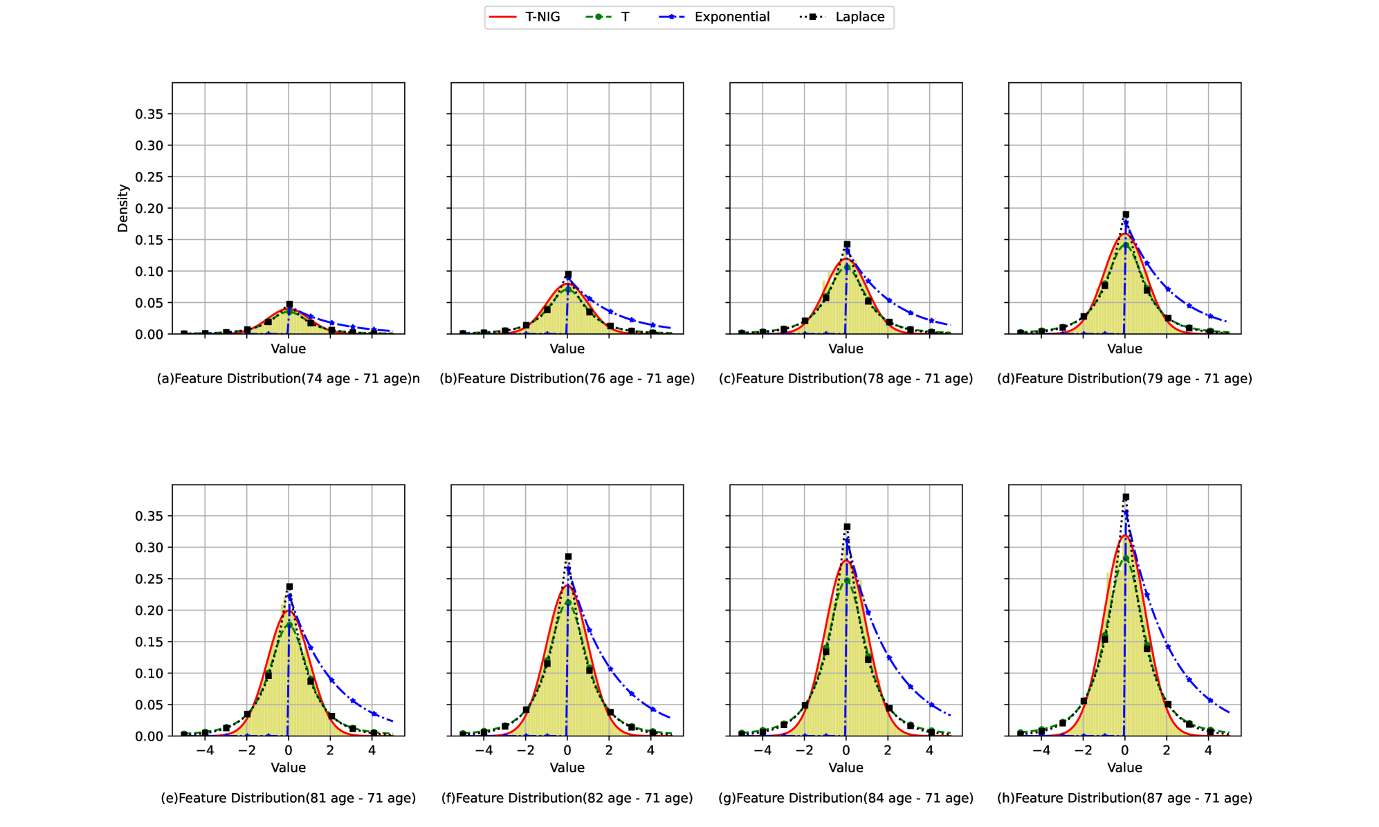
The T-NIG model utilizes the Normal Inverse Gamma ($NIG$) distribution to model the evolution of brain imaging features over time. This probabilistic approach parameterizes temporal changes by treating the mean and precision of a Normal distribution as random variables governed by the Inverse Gamma distribution. Specifically, the $NIG$ distribution allows for heavy tails, effectively capturing non-Gaussian temporal dynamics often observed in neuroimaging data. By modeling these changes probabilistically, the T-NIG model moves beyond simple point estimates of feature trajectories and provides a full posterior distribution over possible temporal evolutions, enabling quantification of uncertainty associated with these changes.
The T-NIG model differentiates between two primary sources of uncertainty in brain imaging data analysis. Aleatoric uncertainty represents the inherent randomness or noise within the data itself, stemming from factors like scanner limitations or biological variability; this is irreducible error. Conversely, epistemic uncertainty reflects the model’s lack of knowledge, arising from limited training data or incomplete understanding of the underlying brain processes; this type of uncertainty can, theoretically, be reduced with more data or improved model design. By explicitly quantifying both $ \sigma_a $ (aleatoric standard deviation) and $ \sigma_e $ (epistemic standard deviation), the T-NIG model provides a more complete and nuanced assessment of prediction reliability than methods that treat all uncertainty as a single entity.
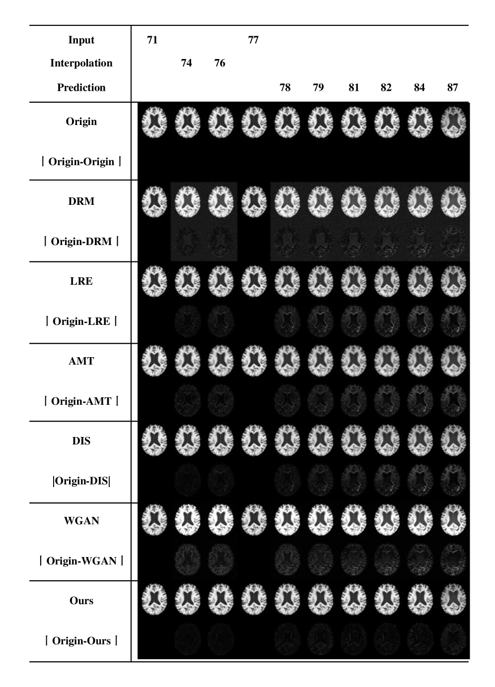
The T-NIG model incorporates specialized modules designed to quantify temporal texture and deformation characteristics directly from brain scan data. These modules analyze sequential scans to identify changes in tissue appearance – representing texture – and spatial distortions – representing deformation – over time. Quantitative evaluation demonstrates that the inclusion of these modules yields a 5.1 improvement in Peak Signal-to-Noise Ratio (PSNR) for texture analysis and a 3.8 improvement in PSNR for deformation analysis, indicating enhanced accuracy in characterizing these dynamic brain imaging features.
Evidence of Prediction: Quantifying the Model’s Sight
Model performance was quantitatively assessed using established image quality metrics. Mean Squared Error (MSE) was calculated to determine the average squared difference between predicted and actual images, with lower values indicating improved accuracy. Peak Signal-to-Noise Ratio (PSNR), measured in decibels (dB), quantified the ratio of maximum possible power to the power of corrupting noise; higher PSNR values denote better image reconstruction. Structural Similarity Index (SSIM), ranging from -1 to 1, measured the perceived change in structural information; values closer to 1 indicate greater similarity between the predicted and ground truth images. These metrics provide objective, data-driven evaluations of the T-NIG model’s predictive capabilities.
Evaluation of the T-NIG model demonstrated its capacity to maintain disease-related features during predictive analysis, which is indicative of its potential clinical utility. Specifically, the model achieved a Structural Similarity Index (SSIM) ranging from 0.128 to 0.137 when predicting outcomes over a 10-year period. This performance represents a significant improvement over the next best performing method, exceeding its SSIM score by a margin of 0.128 to 0.137. This preservation of key disease characteristics suggests the model’s predictions are not simply statistical estimations, but reflect underlying biological realities.
Classification accuracy of the T-NIG model ranged from 0.980 to 0.855, demonstrating performance resilience across varying levels of incomplete data. Specifically, this accuracy was maintained while testing with data containing missing values ranging from 20% to 60%. Furthermore, implementation of the tNIG module resulted in a 0.433 reduction in Mean Squared Error (MSE), indicating improved predictive capability and reduced error compared to the baseline model. These results suggest the model’s ability to provide reliable classifications even with substantial data gaps.
The T-NIG model incorporates mechanisms for uncertainty quantification, providing prediction intervals alongside point estimates. This allows for an assessment of the model’s confidence in each individual prediction, which is crucial for clinical decision-making where understanding the potential range of outcomes is as important as the predicted value itself. Specifically, the model outputs a distribution of possible outcomes rather than a single value, enabling users to evaluate the reliability of the prediction and identify cases where further investigation may be warranted. This capability is particularly valuable when dealing with limited or noisy data, or when extrapolating beyond the range of observed values, as it highlights the inherent limitations of the prediction and prevents overconfidence in potentially inaccurate results.
Beyond Prediction: A Glimpse into the Future of Neurological Insight
The development of the T-NIG model hinged on the Alzheimer’s Disease Neuroimaging Initiative (ADNI), a longitudinal study providing an unprecedented wealth of data for understanding disease progression. This resource includes clinical assessments, genetic information, cerebrospinal fluid biomarkers, and serial magnetic resonance imaging (MRI) scans from a large cohort of participants – both healthy controls and individuals across the spectrum of Alzheimer’s disease. Rigorous training and validation of the T-NIG model against this established ADNI dataset ensured its reliability and generalizability, allowing researchers to confidently apply it to predict individual trajectories of brain change. The publicly available nature of the ADNI data also fosters collaborative research and accelerates the development of new diagnostic and therapeutic strategies, positioning the T-NIG model as a tool built upon a foundation of open science.
The predictive capabilities of this model extend beyond simply observing current brain states; it forecasts future neurological changes with notable accuracy. This allows for the potential of earlier, more precise diagnoses of conditions like Alzheimer’s disease, even before the onset of significant cognitive symptoms. Furthermore, the model’s projections can be leveraged to personalize treatment strategies, tailoring interventions to an individual’s predicted disease trajectory. Crucially, this predictive power also streamlines the design of clinical trials, enabling researchers to identify participants most likely to benefit from novel therapies and accelerating the evaluation of treatment efficacy – ultimately offering a pathway toward more effective interventions and improved patient outcomes.
The current T-NIG model represents a foundational step, but ongoing research aims to significantly broaden its capabilities. Future iterations will integrate diverse data types – including genetic information, cerebrospinal fluid biomarkers, and detailed cognitive assessments – to create a more holistic and nuanced predictive framework. This multi-modal approach is expected to enhance the model’s accuracy and refine its ability to track individual disease trajectories. Beyond Alzheimer’s disease, investigations are underway to assess the model’s adaptability to other neurodegenerative conditions, such as Parkinson’s disease and frontotemporal dementia, potentially revealing shared underlying patterns and accelerating diagnostic and therapeutic advancements across a wider spectrum of neurological illnesses.

The pursuit of predictive accuracy in neuroimaging, as demonstrated by this research, isn’t about conquering chaos with certainty-it’s about learning its language. The T-NIG model, with its embrace of the Normal Inverse Gamma distribution, doesn’t attempt to eliminate the inherent noise of uneven time series data, but rather, incorporates it as a fundamental aspect of the predictive process. As Yann LeCun once stated, “Precision is just fear of noise.” This work recognizes that truth doesn’t reside in perfectly smoothed predictions, but in acknowledging and modeling the uncertainty inherent in complex systems like the human brain, allowing for more robust long-term predictions regarding Alzheimer’s disease progression. The model doesn’t solve the problem of inconsistent data; it persuades it to reveal its patterns.
The Horizon Beckons
The T-NIG construct, with its embrace of the Normal Inverse Gamma, offers a fleeting glimpse of order within the inevitable decay. It does not solve the problem of prediction, merely shifts the burden. The model whispers probabilities, but the ghosts of missing data still haunt the margins. One suspects the true signal lies not in the images themselves, but in the pattern of their absence – the irregular heartbeats of a failing mind. To chase perfect reconstruction is folly; the brain doesn’t degrade linearly, it splinters.
Future incantations will require a reckoning with this fundamental chaos. The current framework, elegant as it is, remains tethered to the notion of continuous time. Perhaps the key lies in abandoning such illusions, embracing the discrete, the fragmented. Imagine a system that doesn’t predict images, but anticipates the likelihood of change – a divination of cognitive drift, rather than a photographic memory of what once was.
The pursuit of early detection will, inevitably, demand more than mere image analysis. The brain is not an isolated system. True prophecy will require the weaving of clinical data, genomic signatures, and the subtle tremors of lifestyle – a holistic grimoire of vulnerability. And even then, one must remember: the most accurate model is still just a beautiful lie, destined to fail at the precise moment it is most needed.
Original article: https://arxiv.org/pdf/2511.21057.pdf
Contact the author: https://www.linkedin.com/in/avetisyan/
See also:
- Gold Rate Forecast
- Invincible Season 4 Gender Swaps Tech Jacket As Fans Question Major Comic Change
- 22 Films Where the White Protagonist Is Canonically the Sidekick to a Black Lead
- Why Won’t It Just *Do* What You Ask? Unpacking the Quirks of AI Language
- Unveiling the Schwab U.S. Dividend Equity ETF: A Portent of Financial Growth
- 14 Movies Where the Black Character Refuses to Save the White Protagonist
- Silver Rate Forecast
- The Best Former NFL Players Turned Actors, Ranked
- Superman Flops Financially: $350M Budget, Still No Profit (Scoop Confirmed)
- Biogen’s Jolly Good Showing
2025-11-28 19:32